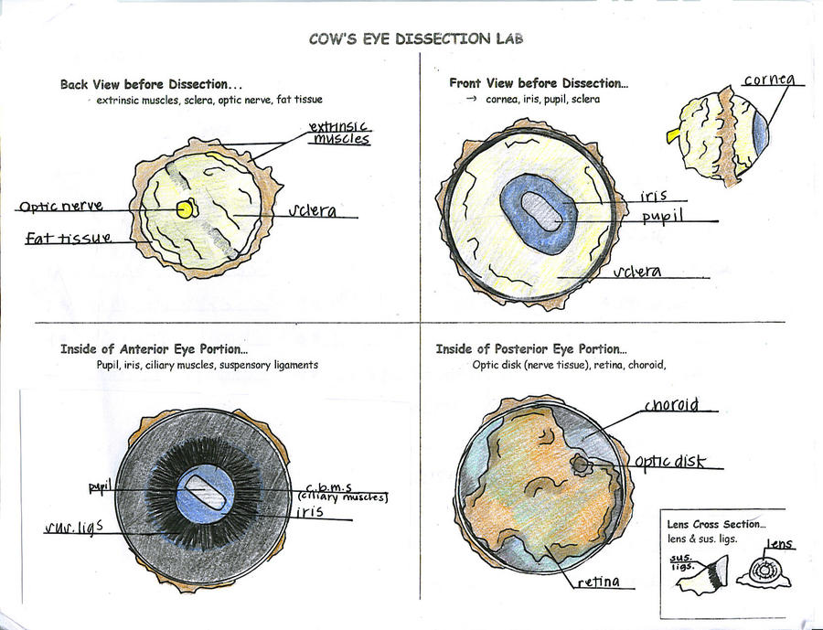
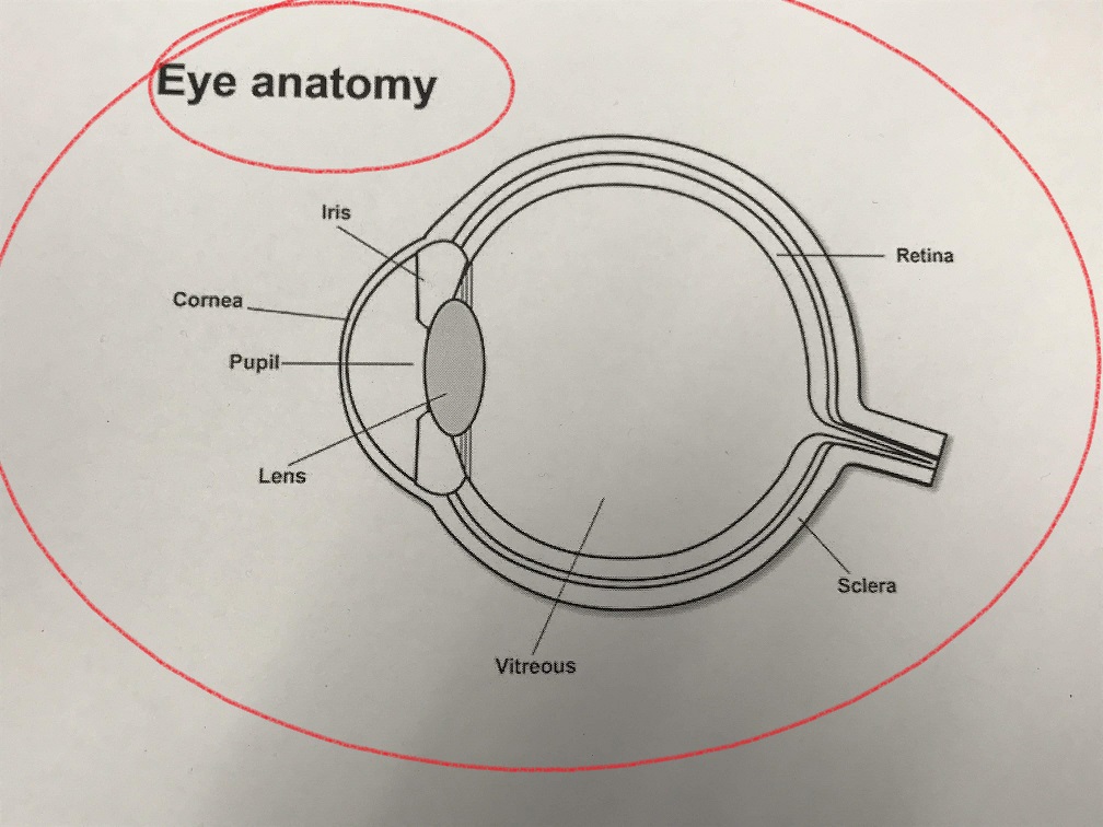
38 labeled cow eye diagram
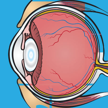
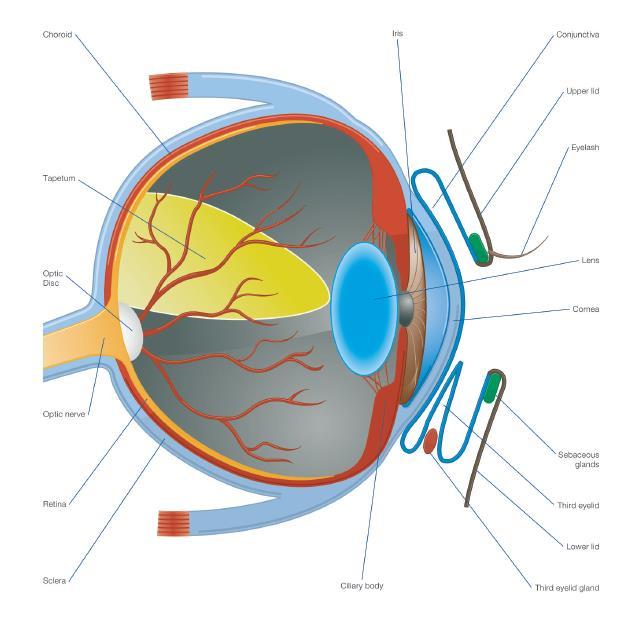
PDF COW'S EYE dissection - Exploratorium This diagram shows the parts of the eye. Can you find these parts in a cow's eye? SCLERA TAPETUM OPTIC NERVE BLIND SPOT LENS VITREOUS HUMOR IRIS CORNEA RETINA AQUEOUS HUMOR PUPIL COW'S EYE dissectionpage 3 Examine the outside of the eye. See how many parts of the eye you can identify. Quantitative analysis of the size of ocular dominance columns. The ... Download scientific diagram | Quantitative analysis of the size of ocular dominance columns. The proportions of labeled area to a given cortical area were measured and plotted for each site. See ...
Cow Anatomy - External Body Parts and Internal Organs with Labeled Diagram The external body parts from the head region of a cow - in this head region, you might identify the mouth, lip, cheek, chin, muzzle, forehead, poll, ear, eye, nostril, and other. Different parts from the neck region of a cow - here, you will find the neck crest, dewlap, brisket, and jugular groove.
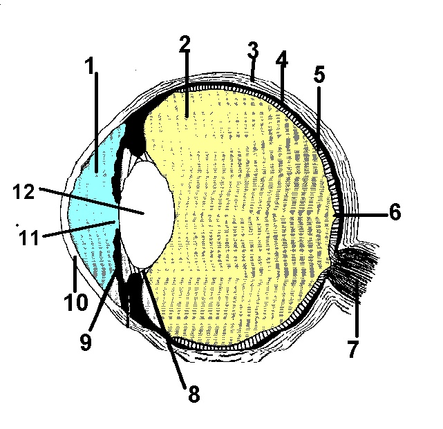
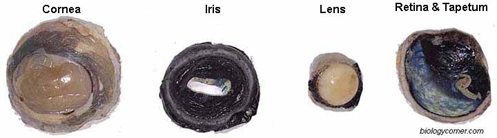
Labeled cow eye diagram
Cow Eye Lab Flashcards | Quizlet beautiful blue-green part of cow eye reflective layer usually located between retina and choroid allows additional light to stimulate the retina nocturnal animals Retina: Photoreceptors light sensing portion of the retina Retina: Photoreceptors--Cones respond to color information primarily clustered around the fovea responsible for center vision Eye Anatomy: Parts of the Eye and How We See The surface of the eye and the inner surface of the eyelids are covered with a clear membrane called the conjunctiva. The layers of the tear film keep the front of the eye lubricated. Tears lubricate the eye and are made up of three layers. These three layers together are called the tear film. The mucous layer is made by the conjunctiva. Cow Eye Dissection & Parts of the Eye Diagram | Quizlet Clear, outer layer of the front of the eye. sclera White, outermost layer of the eye. Helps maintain shape and gives attachment to muscles. photoreceptors The cells in the retina that respond to light (rods and cones) rods Photoreceptor cells in the eye that detect black, white, and gray cones Photoreceptor cells in the eye that detect color
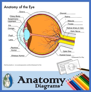
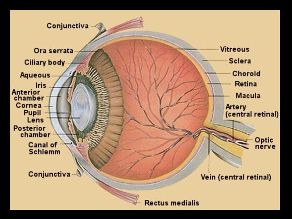
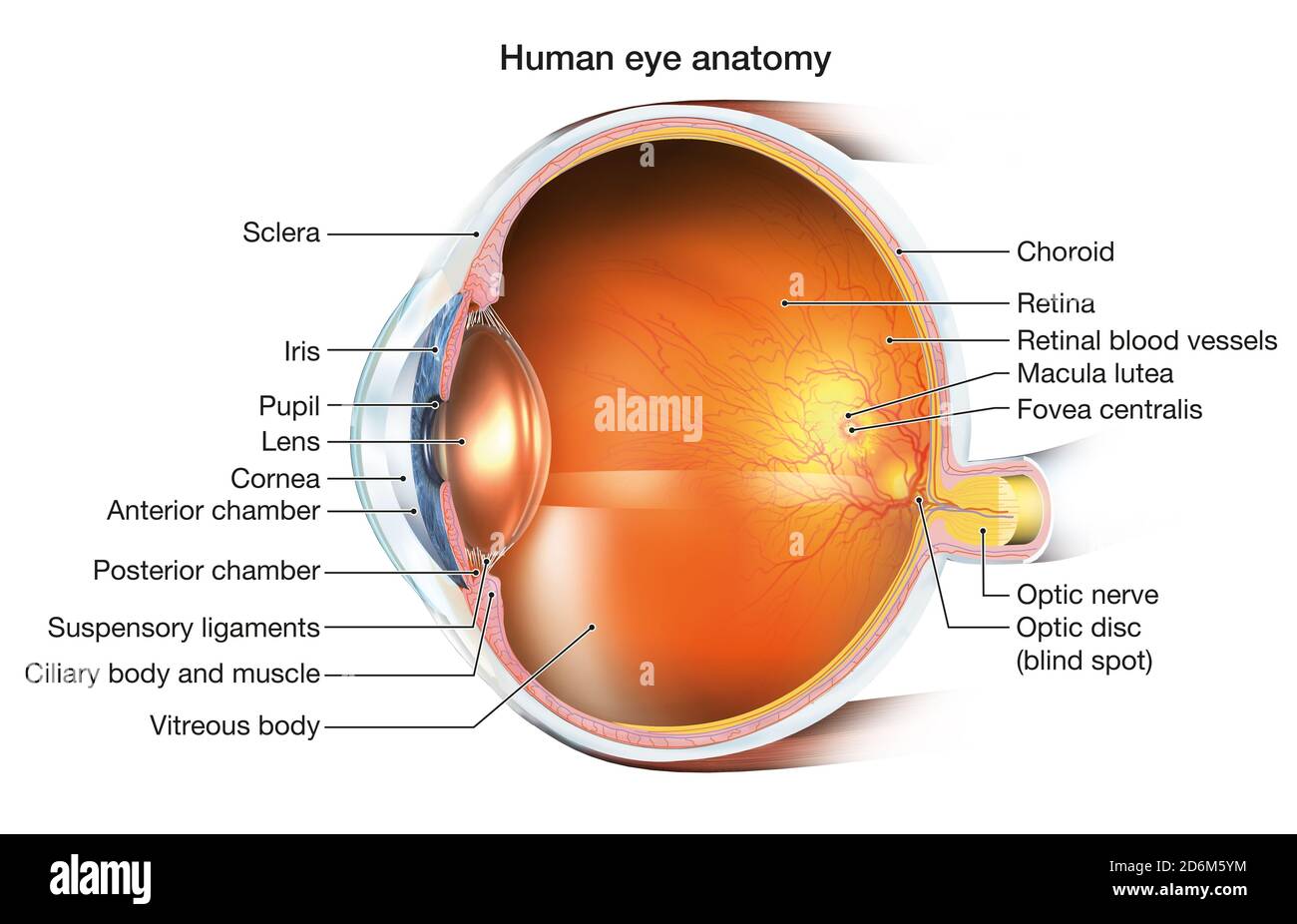
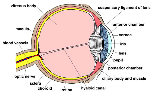
Labeled cow eye diagram. Cow Eye Labeled Diagram - ClipArt Best 31 cow eye labeled diagram. Free cliparts that you can download to you computer and use in your designs. Eye Diagram With Labels and detailed description - BYJU'S A brief description of the eye along with a well-labelled diagram is given below for reference. Well-Labelled Diagram of Eye. The anterior chamber of the eye is the space between the cornea and the iris and is filled with a lubricating fluid, aqueous humour. The vascular layer of the eye, known as the choroid contains the connective tissue. Cow's Eye Diagram Quiz - PurposeGames.com Label the parts of the eye. This online quiz is called Cow's Eye Diagram. It was created by member jenniferbjensen and has 11 questions. The Eyes (Human Anatomy): Diagram, Optic Nerve, Iris, Cornea ... - WebMD The front part (what you see in the mirror) includes: Iris: the colored part. Cornea: a clear dome over the iris. Pupil: the black circular opening in the iris that lets light in. Sclera: the ...
Eye Anatomy - 3D model by MotionCow [5dac474] - Sketchfab Eye Anatomy - 3D model by MotionCow (@MotionCow) [5dac474] Eye Anatomy 3D Model MotionCow 114.6k 98 Triangles: 64.9k Vertices: 51.7k More model information Realistic cross-section of the Eye. This 3D model can be licensed from MotionCow by Educators, 3D Artists and App Developers. Published 6 years ago Science & technology 3D Models eye cross PDF Download Solutions Label The Human Eye Diagram 8.5 by 11 - Inch. Glossy Paper. Thank You. Eye Anatomy Coloring Book - Sep 13 2020 Make the Perfect Gift for All Ages in Any Occasion Who Loves Coloring. Enjoy the Coloring with 50 Illustrations of Human Eye Anatomy. The Human Eye/Ophthalmology Coloring Book provides a means of learning about the structure and function of the Human The origin of thalamic inputs to the FEFsac , SEF , and ... - ResearchGate Download scientific diagram | The origin of thalamic inputs to the FEFsac , SEF , and PMd in the left hemisphere of monkey C5 on coronal sections ( C5-L ) (see Table 1 for fluorescent tracers used ... Cow Eye Dissection Teaching Resources | TPT - TeachersPayTeachers Materials NeededEye model (or reference illustrations of the eye)Cow Eye (s)Scissors & probesTime Required30 minutes for the model and 30+ minutes for the dissection.This is part of a complete unit, Light, Optics, & Color also sold on th Subjects: Biology Grades: 5th - 9th Types: Homeschool Curricula, Laboratory
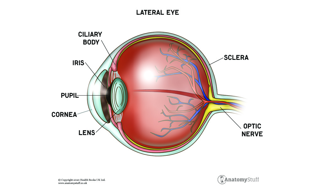
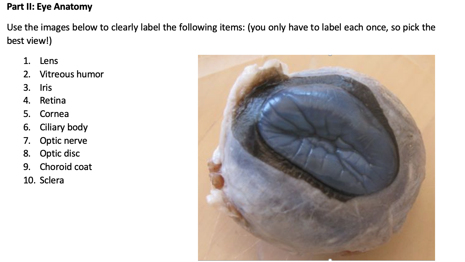
Anatomy of the eye: Quizzes and diagrams | Kenhub How to learn the parts of the eye. Found within two cavities in the skull known as the orbits, the eyes are surrounded by several supporting structures including muscles, vessels, and nerves. There are 7 bones of the orbit, two groups of muscles (intrinsic ocular and extraocular), three layers to the eyeball … and that's just the beginning. Cow Eye Dissection & Anatomy Project | HST Learning Center Look carefully at the preserved cow eye. The most noticeable part of the eye is the large mass of gray tissue that surrounds the posterior(back) of the eye and is attached to the sclera. The second most noticeable part of the eye is the cornea, located in the anterior(front) part of the eye. Anatomy Of Eye Worksheets: Worksheets Cow Eye Dissection Worksheet ... Anatomy Of Eye Worksheets: Worksheets Cow Eye Dissection Worksheet Answers Laurenpsyk Free , Cow's Eye Dissection - Eye diagram - Exploratorium A cow's iris is brown. Human irises come in many colors, including brown, blue, green, and gray. A clear fluid that helps the cornea keep its rounded shape. The pupil is the dark circle in the center of your iris. It's a hole that lets light into the inner eye. Your pupil is round. A cow's pupil is oval.
Cow Eye Dissection | Carolina.com
Bull and the cow - Illustrated atlas : normal anatomy | vet ... - IMAIOS This veterinary anatomical atlas includes 27 scientific illustrations with a selection of labelled structures to understand and discover animal anatomy (skeleton, bones, muscles, joints and viscera). Positional and directional terms are also illustrated.
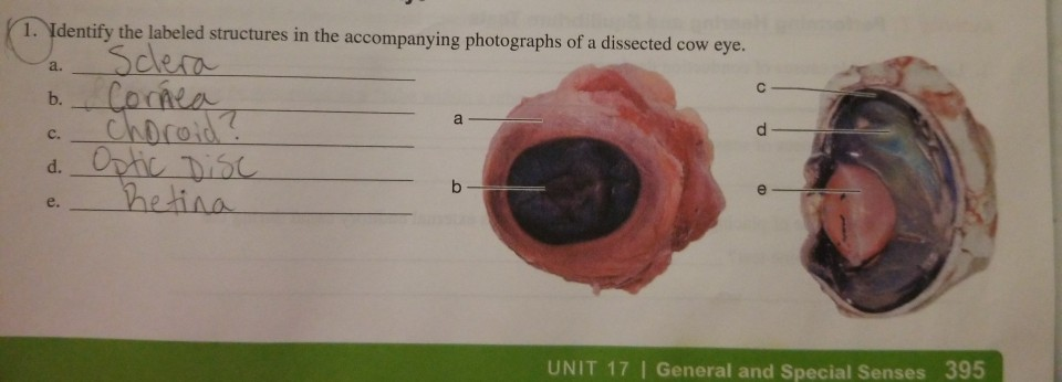
Solved 1. Identify the labeled structures in the | Chegg.com
Cow Eye Dissection & Parts of the Eye Diagram | Quizlet Clear, outer layer of the front of the eye. sclera White, outermost layer of the eye. Helps maintain shape and gives attachment to muscles. photoreceptors The cells in the retina that respond to light (rods and cones) rods Photoreceptor cells in the eye that detect black, white, and gray cones Photoreceptor cells in the eye that detect color

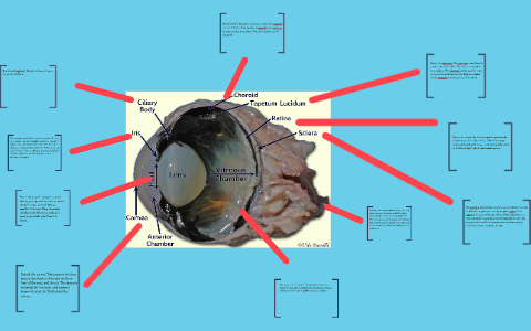
Detailed Cow Eye Dissection: Part II (Jr. High, High School and College Review)
Eye Anatomy: Parts of the Eye and How We See The surface of the eye and the inner surface of the eyelids are covered with a clear membrane called the conjunctiva. The layers of the tear film keep the front of the eye lubricated. Tears lubricate the eye and are made up of three layers. These three layers together are called the tear film. The mucous layer is made by the conjunctiva.
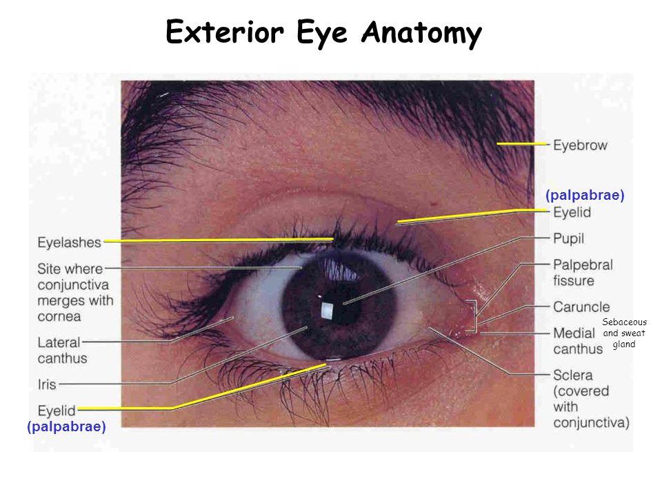
Special Senses Week 12. Exterior Eye Anatomy 1?2? 3? - ppt ...
Cow Eye Lab Flashcards | Quizlet beautiful blue-green part of cow eye reflective layer usually located between retina and choroid allows additional light to stimulate the retina nocturnal animals Retina: Photoreceptors light sensing portion of the retina Retina: Photoreceptors--Cones respond to color information primarily clustered around the fovea responsible for center vision

Human eye Anatomy, Eye, people, orange png | PNGEgg

Muscles of the Human Eye stock illustration. Illustration of ...
Cow Eye Labeled Diagram - ClipArt Best

Blank Eye Diagram - A Useful Resource for Medical Students ...
Development, Anatomy and Physiology of the Eye The word ...
Cow Eye Dissection by tiggerbaby1122 on DeviantArt
The Anatomy of the Cow Eye by Meli C
Eye Anatomy | How does vison work? | Ocular Anatomy

Good Nutrition Can Help Your Eyes | Eye anatomy, Eye anatomy ...
Eye Diagram Teaching Resources | TPT
Anatomy of the Eye Lecture 1 Anatomy of the Eye 1. *The ...
Ciliary body hi-res stock photography and images - Alamy

Figure 31.9 Internal Structures of the Cow Eye Diagram | Quizlet
Sheep Eye Dissection Lab
Solved] Lab 13 (13-11) Label the cow eye. 13. Label the Cow ...

A&P Lab -- Unit 2 -- Labeled Cow Eye Diagram | Quizlet

Cow Eye

Cow's Eye Dissection - Eye diagram
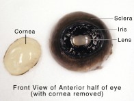
Cow Eye Dissection & Anatomy Project | HST Learning Center

Cow eye – dissection and label
Development, Anatomy and Physiology of the Eye The word ...

Cow Eye Diagram | Quizlet

External and internal anatomy of the cow eye Diagram | Quizlet
Anatomy of the eye
Anatomy of the eye | Children's Wisconsin

2.4.1.A EyeAnatomy SRS.docx - Activity 2.4.1: Exploring the ...
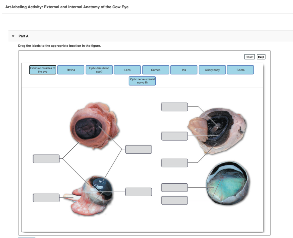
Solved Art-labeling Activity: External and Internal Anatomy ...
COW EYEBALL ANATOMY – NAME THE PARTS
Development, Anatomy and Physiology of the Eye The word ...
Solved Please label each diagram with the appropriate | Chegg.com

Cow Eye Dissection & Parts of the Eye Diagram | Quizlet
Hamburg CSD - 5th Grade Cow Eyes
Cow Eye Dissection
sheep eye dissection procedures
Post a Comment for "38 labeled cow eye diagram"